Deep Sea Fish and Sediment Surveys in the Gulf
Meet C-IMAGE Scientist: Patrick Schwing
Dr. Patrick Schwing is working after graduation as a Postdoctoral scientist for the C-IMAGE project at USF’s College of Marine Science. Enjoy the following Photo Story of his science research and activities during the C-IMAGE August 2012 cruises.
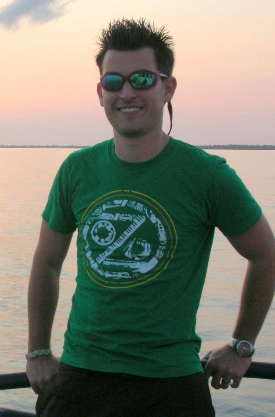
C-IMAGE Scientist: Patrick Schwing (Postdoctoral Researcher)
This is a picture that was taken aboard the R/V bellows during one of our seafloor surveying cruises with Dr. Stan Locker in the Dry Tortugas National Park. We were using a C3D side-scan sonar instrument to obtain a high definition map of seafloor bathymetry. This was paired with airborn LIDAR maps to create a complete map of the National Park.
Following the Deepwater Horizon Blowout in 2010, several cruises went out to assess the impact on the seafloor. This is a picture of the MC800 multicorer that was used to obtain short sediment cores throughout the northeastern Gulf of Mexico. The multicore “lunar lander” is the perfect instrument for capturing the sediment-water interface and surface sediments. It also collects eight cores at once, allowing for several different analyses to be done at the same site with only one cast.
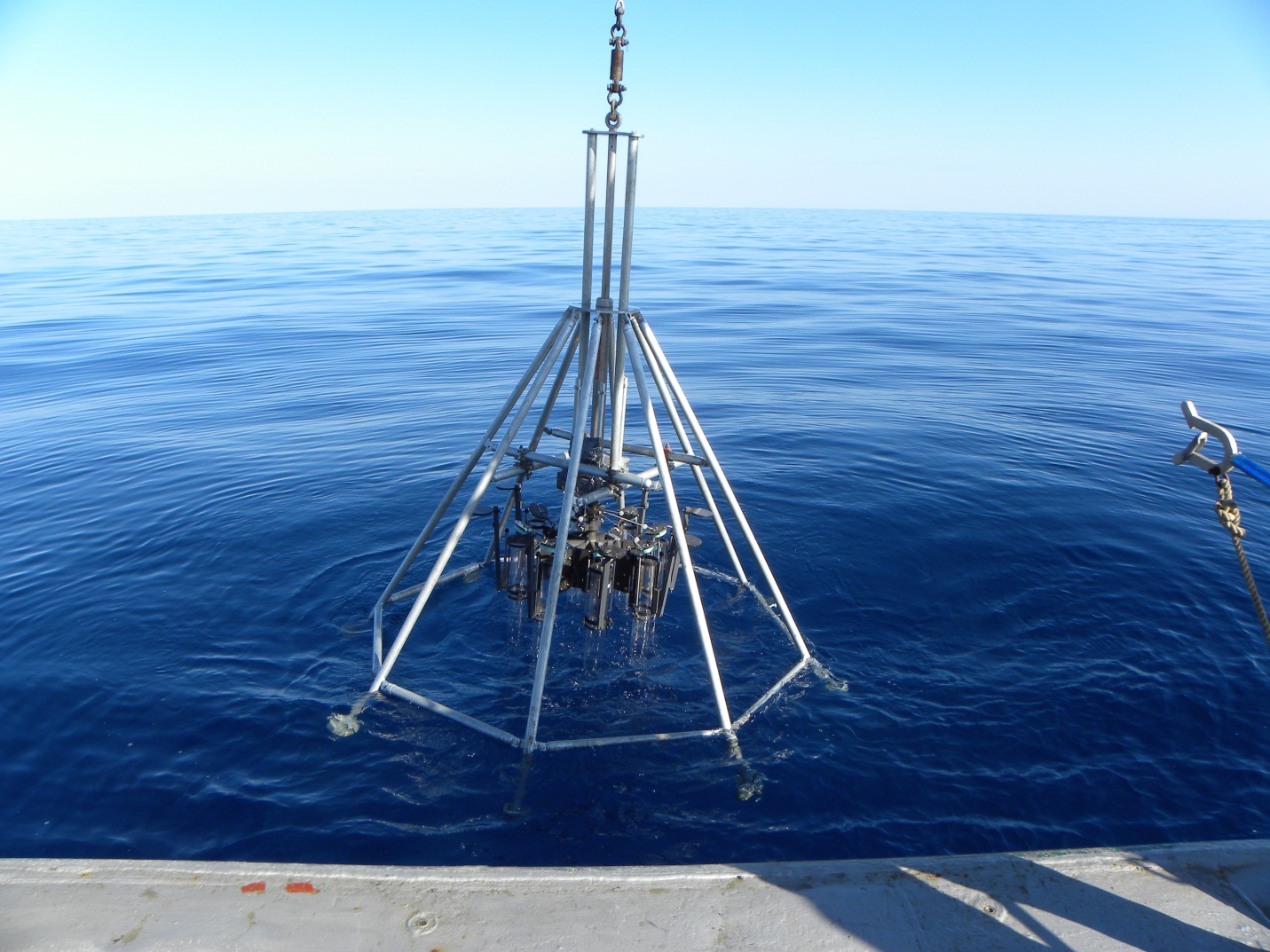
Multicore Sediment Sampler used during C-IMAGE August 2012 cruises
Once the multicore returns to the deck, the sediment tubes must be retrieved as shown below. The sediment is then transferred into other tubes that have been cut to size for each core. They are then sealed and kept at cold temperatures while they await sampling and analysis in the lab.
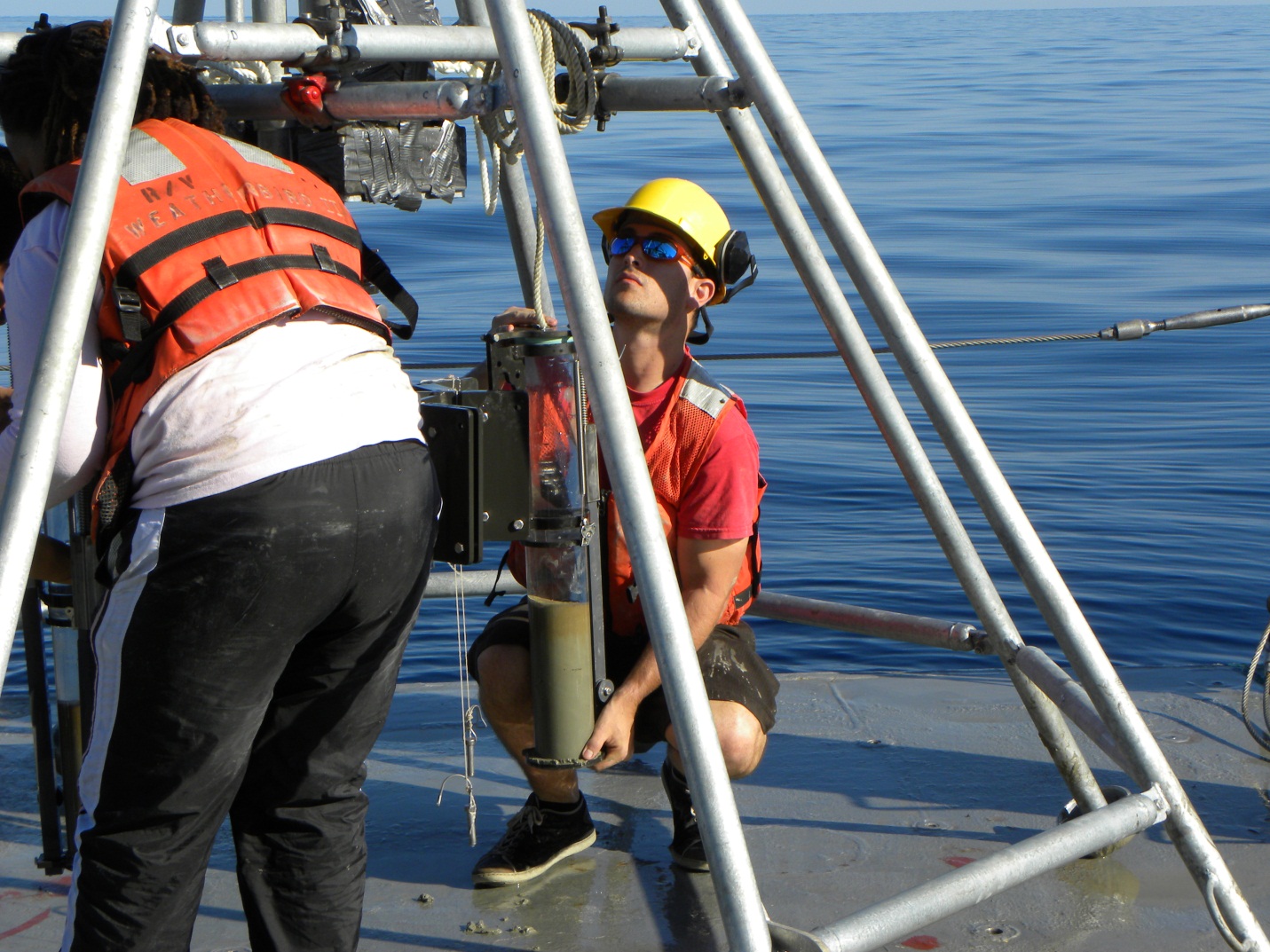
Retrieving sediment samples from seafloor during C-IMAGE cruise
One of the cores is split longitudinally and cleaned for photography. The other seven cores are used for short-lived radioisotope dating, organic geochemistry, benthic foraminifera abundance and stable isotopes, microbial analysis, reduction/oxidation metal chemistry, pore water chemistry, and an archive core.
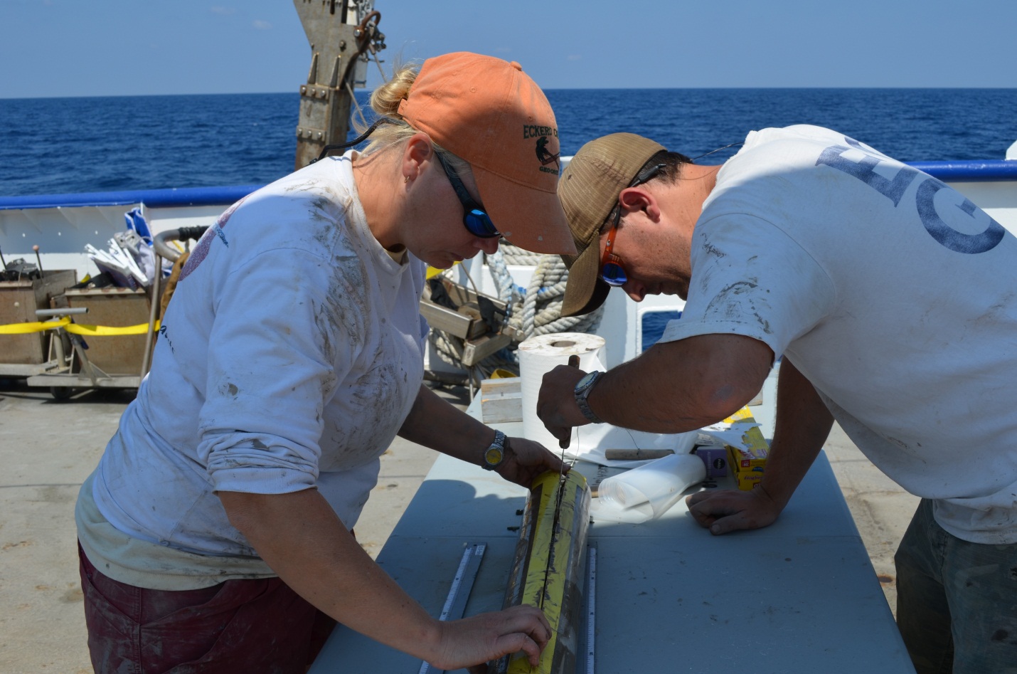
C-IMAGE scientists preparing core for splitting and visual description
This series of photos was taken of three cores taken from the same site at different time intervals. It is evident that a large amount of material has been deposited between August 2010 and February 2011. The short lived radioisotope geochronology has shown that the mass accumulation rates have increased by one, and in some cases, two orders of magnitude. The increase in deposition is most likely due to settling of plankton due to hydrocarbon toxicity or blooms of microbes in the water column that thrived during the blowout.
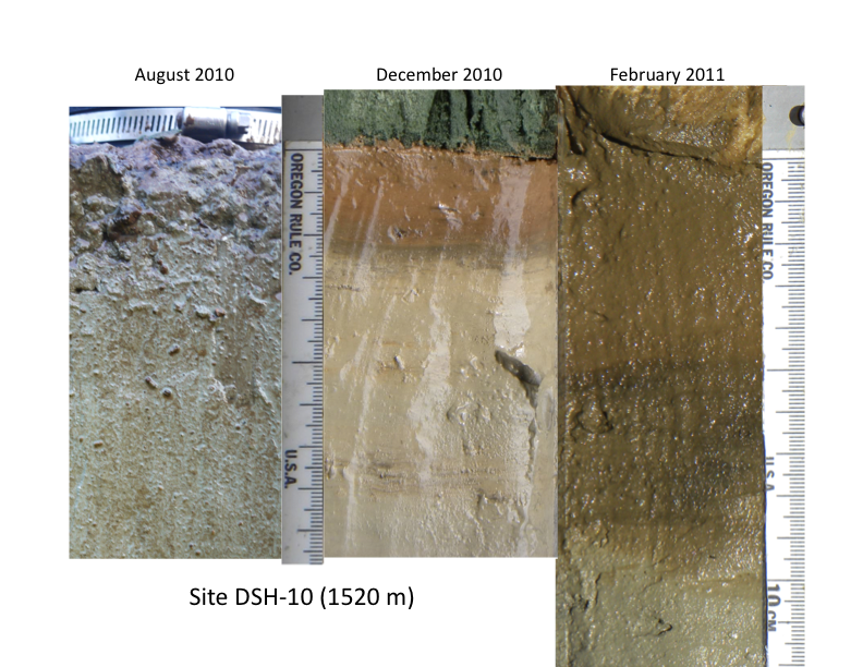
Series of photos from three cores taken from the same site at different time intervals
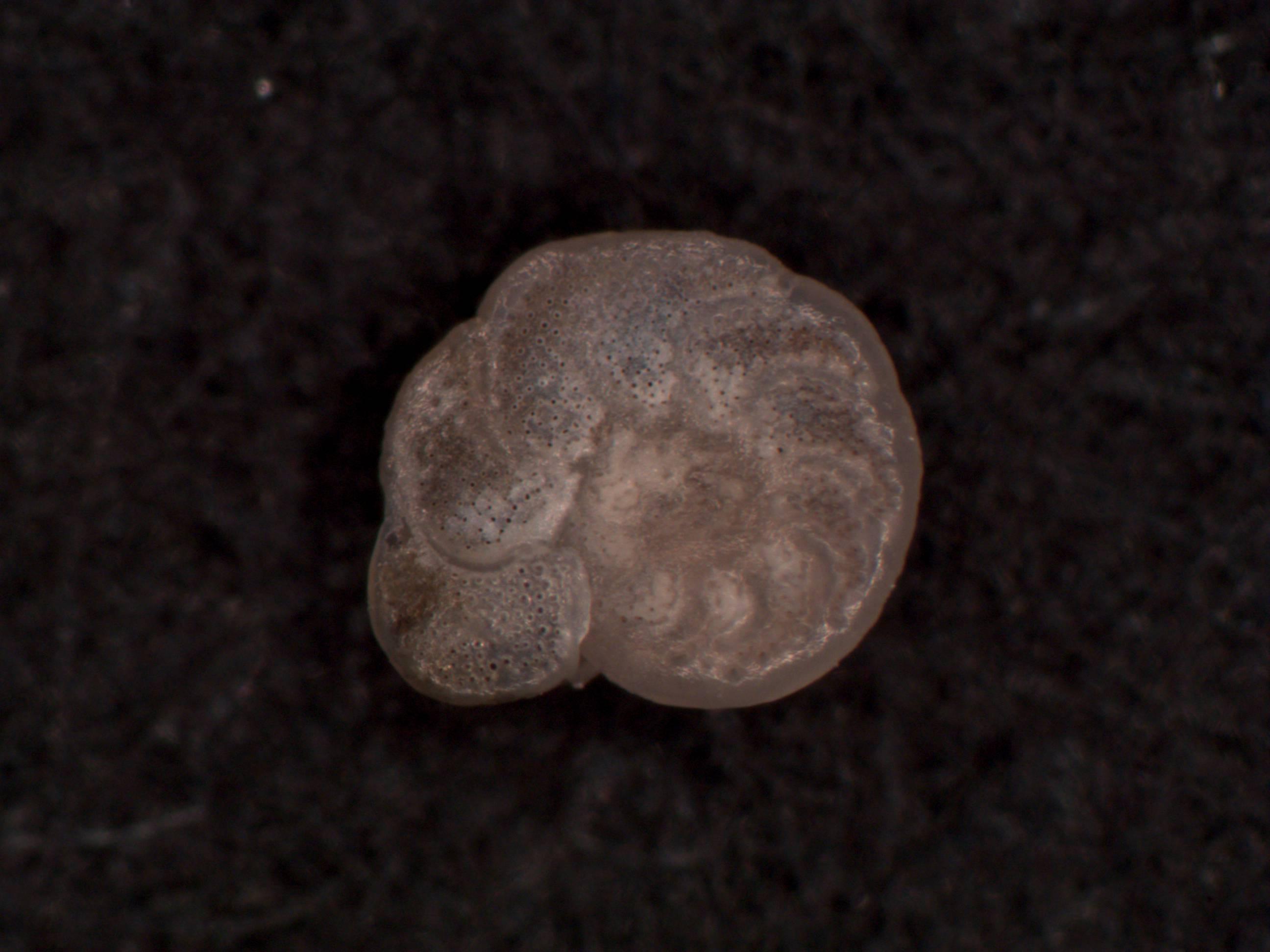
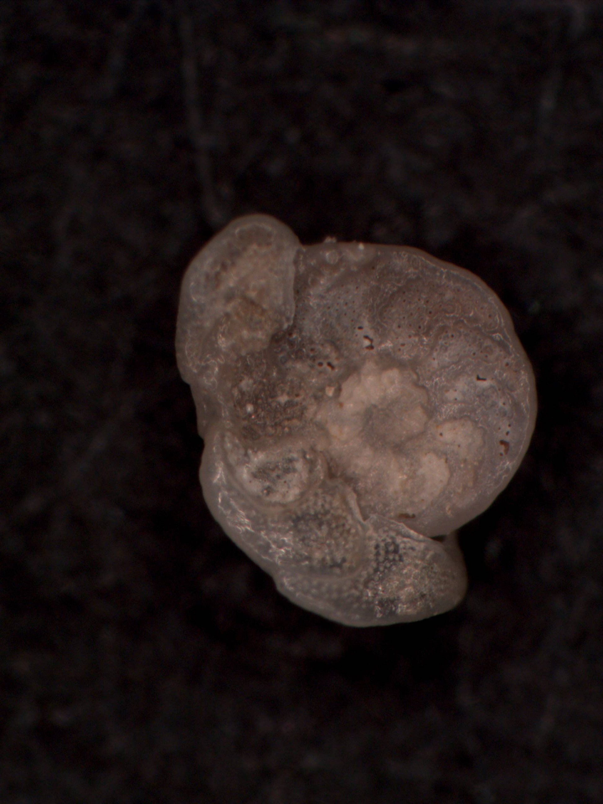
The benthic foraminifera (single-celled carbonate protists) picked from each sample depth in the core were counted, photographed in case of deformation (photo on right), and analyzed for their carbon isotope composition as an indicator of direct contact with hydrocarbons. The foraminifera pictured here are Cibicidoides wuellerstorfi (“healthy” specimen in photo on left).
| Print article | This entry was posted by greely on August 18, 2012 at 10:30 pm, and is filed under Oceanic Updates. Follow any responses to this post through RSS 2.0. You can leave a response or trackback from your own site. |